It would be interesting to take a poll of non-neuroscientists on whether or not they are aware of glia. I’m guessing <5% of people know they are the other half of the ~170 billion cells in your brain (besides neurons). I for one had never heard of them until I took my first (and only, actually) neuroscience course. And it turns out that since Prof. Lubischer (whose splendid class helped further my interest in neuroscience) studied glia, she gave us a running list of all the jobs they performed in the brain. It’s probably a bit outdated 5 years later, but for those interested I put all 12 of them at the end of this post.
I think the main reason hardly anyone knows about glia is that they aren’t so easy to study. Unlike neurons, which scream to be measured with their significant and persistent electrical discharges, glia act largely in a support role—almost like little helper robots to the master neurons. Their main functions involve regulating ions, neurotransmitter levels and therefore the electrical signals of neurons themselves at various key points (e.g. along the neuronal axon, at the synapses between neurons). And since these little chemists work with ~attoliter amounts of product, measuring their effects is tricky.
Now, for the crazy, mad scientist stuff I hinted at in the title. Despite their passive role relative to neurons, glia potentially can have effects on a cognitive level. In a paper published this year in Cell Stem Cell, a group of neuroscientists led by Stephen Goldman and Maiken Nedergaard at the U. of Rochester Medical Center took human progenitor glial cells (straight from ~5 month old aborted fetuses) and grafted them into the brain of immunodeficient (I presume so the mouse brain wouldn’t attack the invading human glia) baby mice. Humans have notably larger and, frankly, better glia than mice when it comes to their role in regulating ions and speeding up neurotransmission. Therefore, they probably went in thinking the human glia might alter the performance of the native mouse neurons.
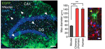
The left picture below shows some of these human glia (in green; the nuclei of human glia are in white) that were incorporated into my favorite part of the brain: the hippocampus. The authors note the human cells—after 14 months in the mouse brain—are particularly enriched in the dentate gyrus region of the mouse hippocampus. Dentate gyrus granule neurons are the strong c-shaped band of blue cells in the left figure. These neurons are one of only two groups that can be newly born in mammalian adults and are heavily implicated in memory and emotions (I study this region in primates as it is also thought to be an essential brain region for how we separately encode new memories). Human glia also were expressed to a lesser degree throughout the mouse cortex (the cortex is kind of like a heating helmet for the important hippocampus underneath—haha I’m just kidding. ALL of the brain is important).
(from http://dx.doi.org/10.1016/j.stem.2012.12.015)In the right picture are a few of these human glia (green) compared to the native mouse glia (red arrows)—specifically, astrocytes. As you can see, this subclass of glia are called this because of their distinctive star-like shape. I’ll largely refer to the human glia as human astrocytes from here on since the authors used this obvious morphology to select for glial progenitor cells that had ‘grown up’ within the mouse brain into developed astrocytes. And when these human astrocytes did grow up, they managed to grow to the size regular astrocytes do in the human brain—much bigger than the native mouse ones (see middle graph). This confirmed previous work that had shown how human glia incorporated themselves into mouse brains, performed a usual glia task as I explained above (in this previous case—insulating ‘leaky’ axons to heal congenitally deformed mice), and maintained their typical size and morphology in mouse brains as normally seen in human brains. Basically: the human astrocytes are like the honey badger—they don’t care what brain they’re in, they’re gonna go regulate some neurons! Already kinda sweet, right?
Well it gets better. A quick overview of some nitty-gritty details: the authors then went about seeing if there were any biophysical changes in the human vs. mouse astrocytes by studying slices from the postmortem chimeric mouse hippocampus (a common in vitro method). Indeed, they found waves of calcium (an important ion that astrocytes use to regulate neuronal transmission) were 3x faster in the human glia compared to the mouse glia. They also found excitatory postsynaptic potentials (EPSPs) were stronger in the chimeric brains. Further, long-term potentiation (LTP)—a commonly measured signal in the hippocampus that is important in learning and forming memories through the strengthening of neuronal connections—was enriched in the mice with human astrocytes. The authors were even able to show how the human astrocytes specifically did it: by releasing a chemical called TNFα that told the neurons to make more excitatory receptors (the place where neurotransmitters have their effect—more receptors equals easier excitation, hence the boost in LTP). The human astrocytes essentially were able to improve the ability of the mouse brain to transmit signals.
So, you can probably guess what’s next: test these super-brained mice on some typical mousey tasks and see if their more potent brains lead to cognitive enhancements. And amazingly: they did! Chimeric mice showed better memory for a context they had previously been shocked in vs. a similar one where they had not: indicative of a learned, hippocampal memory. In another test of hippocampally-dependent learning, the chimeric mice achieved greater success in the Barnes maze (where the mice must remember the location of a small, dark escape hole–mice hate open, well-lighted places. And Hemingway.). Finally, a third test of hippocampal learning showed the chimeric mice were better able to recognize a familiar object in a novel location. These three tasks might not sound that exciting to prove your supermouse’s worth, but they’re standard tests that depend on the hippocampus. Mice with lesioned hippocampi perform worse on such tasks. Interestingly, these tests are similar to those used to screen for antidepressants (that often then work when given to humans—we’re not so concerned about mouse depression) as the hippocampus is also tied into emotional centers of the brain.
Crazy/sexy/cool right? Okay maybe just 1&3. But what does this mean for you, science-interested human that made it this far? For one thing, we can no longer just think of glia as passive, boring helpers. They’re more like strong lobbyists that may be essential for our cognitive function! And the fact that all it took to enhance the learning and memory of mice was the strategic implementation of one chemical (TNFα), we can now start to target both this cytokine and glial functionality in disorders of cognitive processes. Even better, Stephen Goldman’s lab has been able to induce pluripotent human stem cells from skin cells. This not only eliminates the need for fetal stem cells, but also allows for stem cells already tailored for a specific person to be created (using foreign stem cells could cause an immune response; hence the use of immunodeficient mice in this study). His lab is reportedly already using this system to study mouse models of schizophrenia and Huntington’s, and I’d bet you a greenback that Alzheimer’s won’t be far behind. Depression seems like an awesome candidate for glial therapy as well. Practically no new class of antidepressants has been made for 50 years—most work has concentrated on the same few monoamines in the same neuronal pathway. But it is possible that glia could be used to modulate any number of factors, and do so with the attoscale specificity that popping in a pill of Prozac lacks.
Finally, as a fun aside, this isn’t the first time glia have been implicated in improving cognitive function. Specifically, Marian Diamond’s lab analyzed tissue from Albert Einstein’s brain and found he had an enhanced number of glia compared to an average person. This study was done a while ago and is under some debate (and is possibly not the only abnormality in Einstein’s brain), but it makes for a fun story. Plus, when research started indicating glia could send long-distance chemical signals to each other, their potential role in cognitive functions was no longer that crazy. I, for one, welcome our new glia overlords. And can’t wait to hear about more glial therapies.
List of glial functions:
Glia can…
…regulate the extracellular milieu (e.g. potassium buffering).
…influence axonal propagation through myelin.
…direct localization of membrane proteins.
…uptake neurotransmitters at synapses.
…express neurotransmitter receptors and directly modulate synaptic transmission.
…synthesize neurosteroids.
…provide guidance for migrating neuroblasts (specifically radial glia).
…can provide chemical clues for axonal pathfinding.
…can modulate synaptogenesis.
…make trophic factors for neurons.
…mediate some forms of synaptic plasticity.
…influence the neuronal response to injury and disease.
